Quantitative medical imaging, body composition, AI-assisted learning, and radiology education.
01C2—T1QUANTITATIVE IMAGING
The Spine Journal Peer-reviewed research
1,000-patient cervical CT morphometry
A study of cervical neuroforaminal dimensions: the spaces through which spinal nerves leave the neck. This work uses a large CT dataset to describe normal anatomical variation.
Morphometric analysis of cervical neuroforaminal dimensions from C2–T1 using computed tomography of 1,000 patients.
Research on lumbar neuroforaminal dimensions measured across plain film radiography, CT, and MRI, examining how measurements compare between imaging methods.
Normative Measurements of L1 to S1 Neuroforaminal Dimensions Derived From Plain Film Radiography, Computed Tomography, and Magnetic Resonance Imaging.
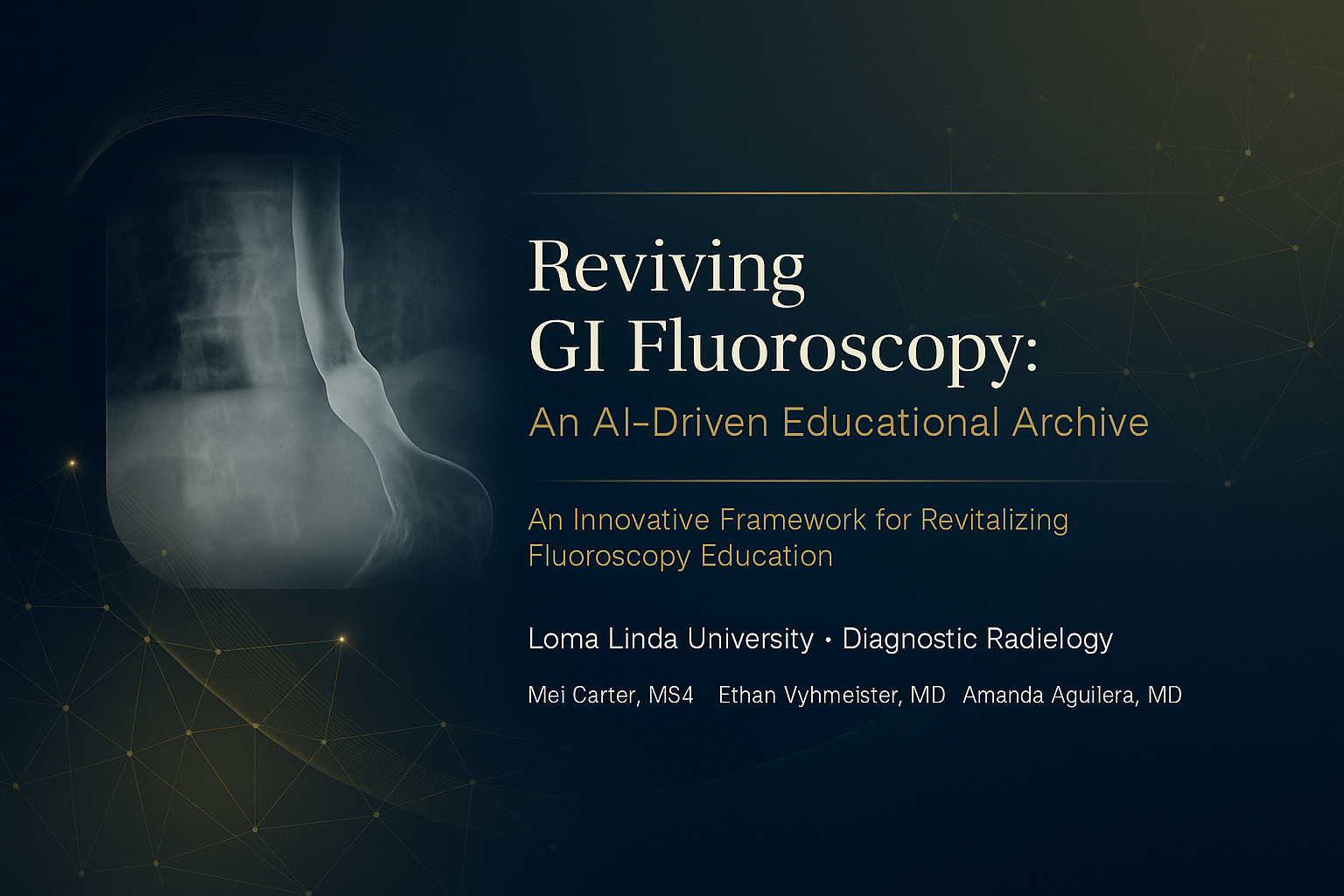
An AI-assisted GI fluoroscopy education platform developed around a large teaching archive. The project brings together radiology education, case-based learning, and practical uses of AI.
Ongoing body-composition research includes centenarian imaging and the study of pectoralis muscle measurements in older adults undergoing elective cardiac surgery. The focus is on muscle area, attenuation, and their relationship with clinical outcomes.
Current research interests: sarcopenia, body composition, aging, and perioperative outcomes.